活動單位 Department
講題
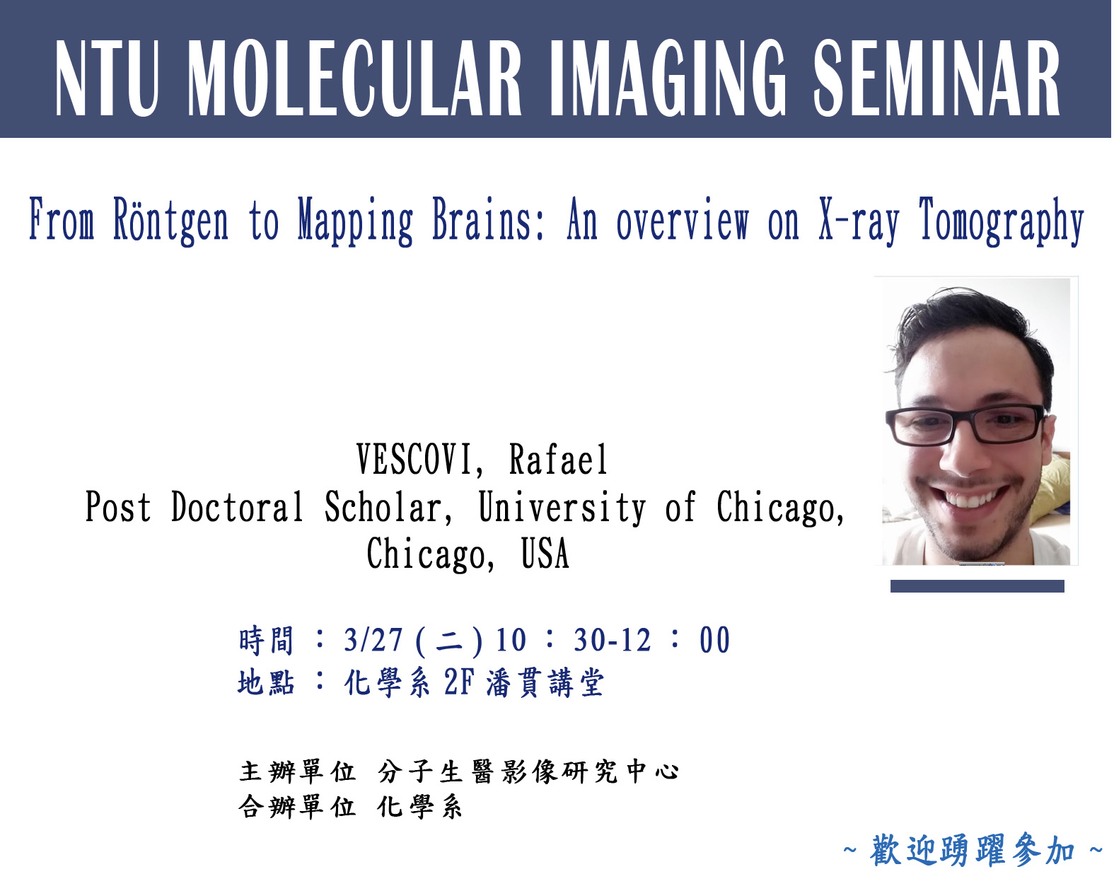
From Röntgen to Mapping Brains: An overview on X-ray Tomography
講者
VESCOVI, Rafael��
Post Doctoral Scholar, University of Chicago, Chicago, USA
時間
3/27(二)10:30-12:00
地點
化學系2F潘貫講堂

內容 Content
Abstract
In 1895 german physicist Wilhelm Röntgen created the first and most famous radiography using x-ray radiation which was unknown at the time. From his systematic study of this new radiation it was cunned the x-ray or "X-rays" (signifying an unknown quantity) though many others referred to these as "Röntgen rays". Since then, x-ray sources have evolved to the point where we can create very bright x-rays from particle accelerators and speed up the use of new techniques that would require immense time to image with lab source x-rays. For biology one of the most common techniques using x-ray is synchrotron X-ray tomographic microscopy (SRXTM). This technique allows for detailed three dimensional revelation of a range of objects with varying spatial resolution. This technique was mainly used in cases where the objects and the surrounding tissues have different densities, like bone and soft tissues but recent tissue preparation techniques stained this piece of tissue with heavy metals without destroying their structure. The last part of this presentation will show how to use SRXTM to probe the cell morphology, blood vessels, dentrites and mielinated axons over whole mouse brains and the prospects to do humans brains.


 繁體中文
繁體中文 English (UK)