PI

Chung-Ming Chen, Professor, Institute of Biomedical Engineering, National Taiwan University
Co-PI
Chiun-Sheng Huang, Professor; Tung-Wu Lu, Professor
The uniqueness of the IR imaging lies in its capability in revealing such metabolic information as heat, oxygenation, and so on, which may not be easily provided by other medical imaging modalities. With the unique strength of the IR imaging, the IR imaging core aims to develop novel low-cost, non-invasive and hazardless multi-spectrum IR imaging technologies to offer effective alternatives to characterize and detect the anatomic and functional disorders of various diseases and changes in tumor growth and treatment response.
A Quantitative Dual-Spectrum IR(QDS-IR) System for Early Detection and Monitoring Chemotherapy Response of Breast Cancer
The QDS-IR provides new tissue parameters characterizing the functional changes in tumor growth and chemotherapy response. The unique idea is to decompose the heat energy on the body surface into those from the high- and normal-temperature tissues with the environmental and physiological influences minimized. With 60 subjects, the [test] of QDS-IR is shown to have the similar variation tendency as the SUVmax of FDG-PET in more than 90% of subjects.
QDS-IR Spectrogram Imaging System
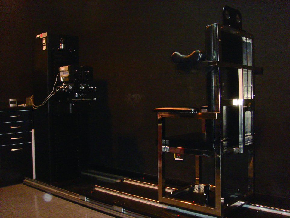
Figure 1. The system architecture of QDS-IR
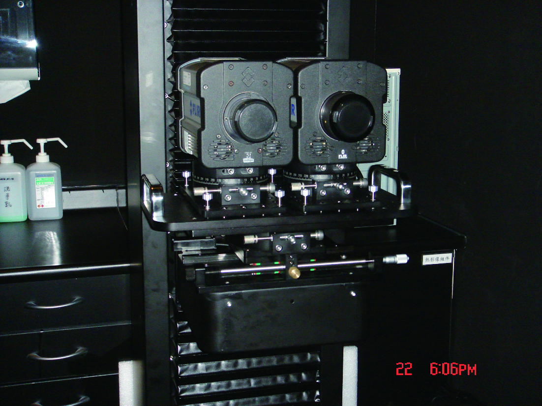
Figure 2. The dual-spectrum IR camera
A Touch-free Infrared thermographic system for Real-time Remote-Monitoring of Free Flap Pedicle Thrombosis
To detect the free flap pedicle thrombosis, a new technique based on the Structural Equation Modeling(SEM) has been developed to reveal the temperature change by eliminating the effect of the latent factors. For the clinical study, the SEM-retrieved specific temperature of the flap demonstrates an evident temperature drop 80 minutes earlier than the clinical finding.
An IR Descriptor for Quantifying The Treatment Response of Complicated Skin And Soft Tissue Infection(cSSTI)
An effective descriptor, denoted by [test], has been proposed for treatment response assessment of cSSTI using IR Images. The uniqueness of the [test] lies in its capability to reveal the relative responses along the course of the treatment. Furthermore, the variation of the [test] was found to be closely related to the phenotype of the MR imaging in the course of cSSTI treatment.
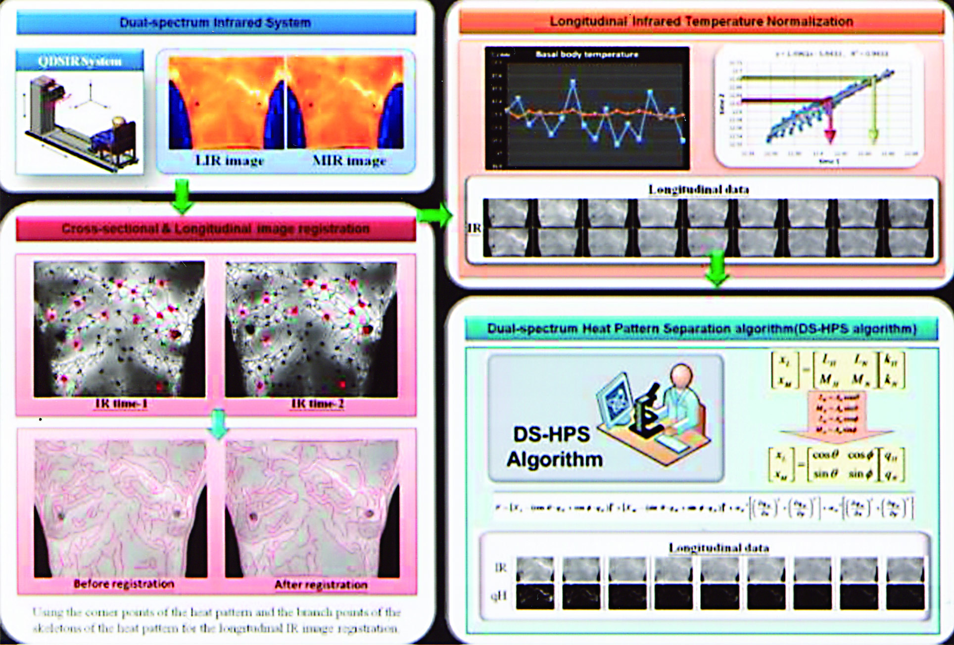
Figure 3. The algorithmic flowchart of the QDS-IR system
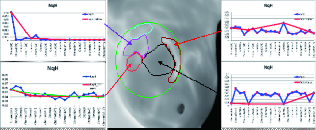
Figure 4. Automatic identification of those regions with different qH tendency along breast chemotherapy
Vessel Thrombosis
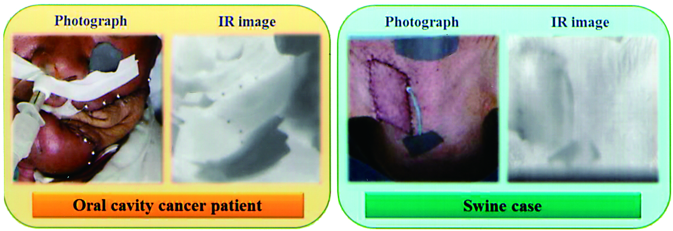
Figure 5. The photos and IR images of the free flap regions of an oral cavity cancer patient and a swine


 繁體中文
繁體中文 English (UK)