Research

2016-12-21 03:43:51 / 核心實驗室


PI

Wen-Yih Isaac Tseng, Professor, Center for Optoelectronic Medicine,

National Taiwan University College of Medicine

CO-PI

Chun-Hung Chen, Professor; Chung-Ming Chen, Professor; Argon Chen, Professor; Hwang-Ching Tai, Assistant Professor  

LAB WEBSITE

http://abmri.mc.ntu.edu.tw/tw/


The mission of digital library core is to develop extensive analysis, modeling, and prediction capability powered by state-of-the-art methodologies, in addition to a traditional library used as a database. Our goal is to serve as both a biological data sharing center, and prediction and decision support system, and to enhance the discovery and availability of molecular imaging biomarkers. Our ultimate goal is to promote early life-threatening diseases detection and prevention, and reduce the mortality rate.

Research facility of digital library core is a hosted Private Cloud System donated by Inventec Inc. and the Institute for Information Industry. The system is installed and managed by the NTU Computer and Information Networking Center. It serves as a platform of data archiving, transferring and computing developed by the digital library core.

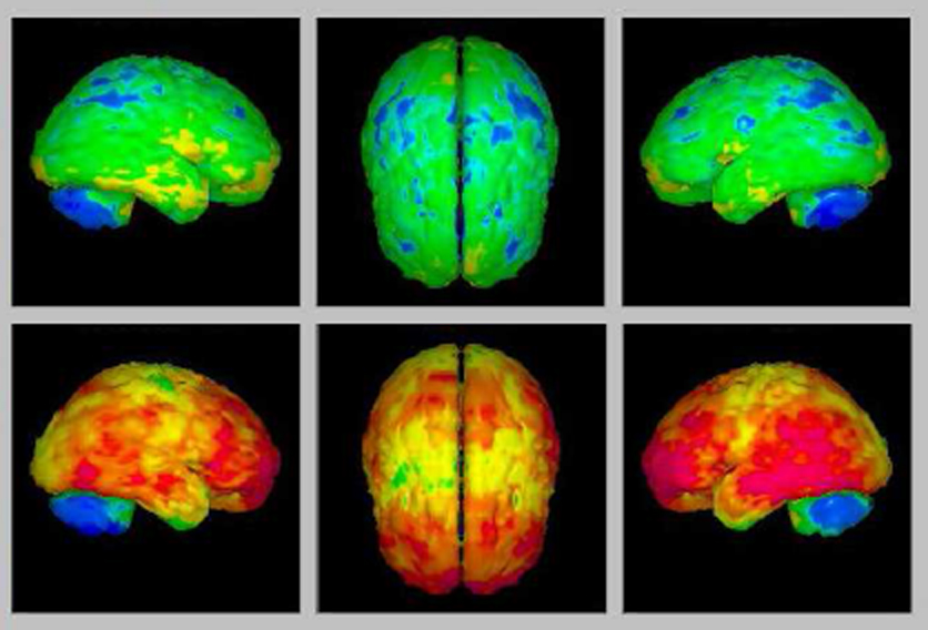
Current progress in the core includes a tractatlas that provides anatomical locations of 74 major white matter tract bundles of the human brain(Figure 1.) and a tract-based automatic analysis pipeline that allows automatic registration of grain images and measuring connection integrity along each tract(Figure 2.). The automatic pipeline allows high throughput analysis of whole-brain white matter tracts in a large cohort of patients. It is applicable to neuropsychiatric diseases, such as schizophrenia, autism spectrum disorder, attention deficit hyperactivity disorder, epilepsy, and Alzheimer's disease, to detect the altered connectivity of the neural network in patients. Currently, we are working on a cloud-based computing platform to provide such unique service through the cloud.

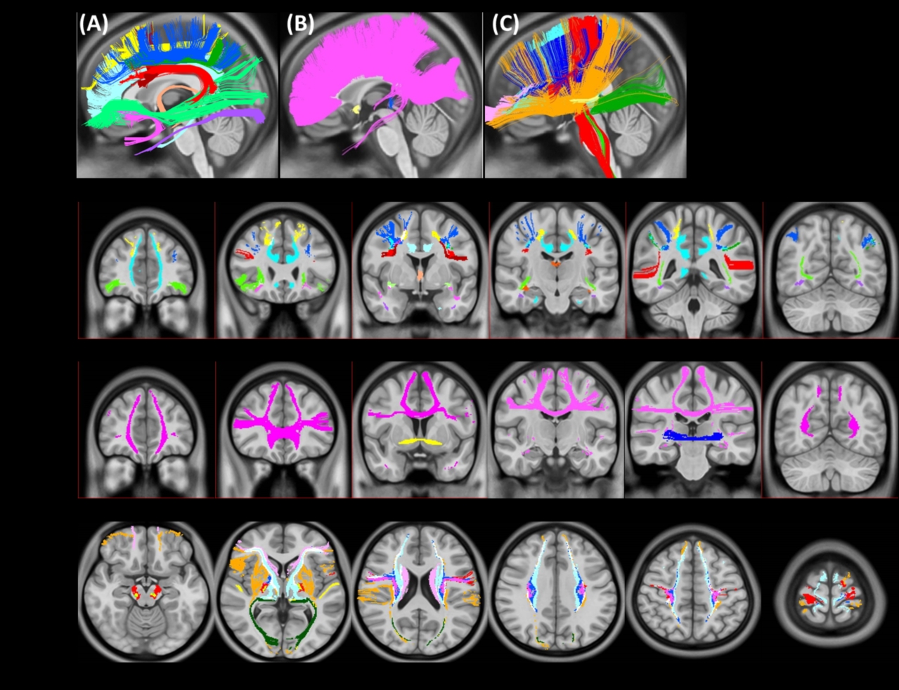
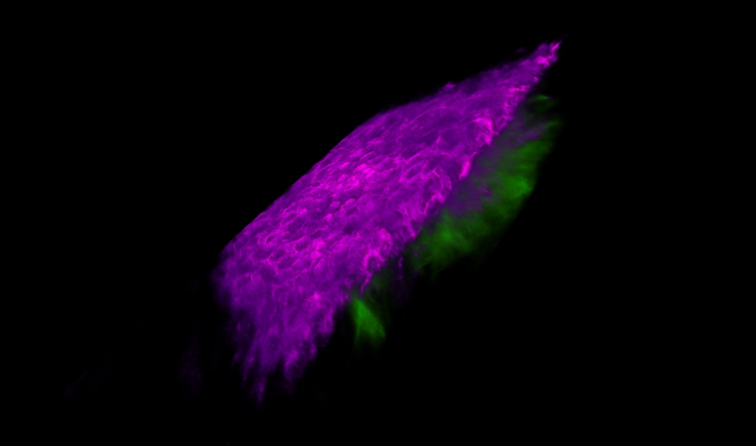
Figure 1. Tractatls provides anatomical locations of 74 major white matter tract bundles of the human brain


Digital Library Core Photo 02

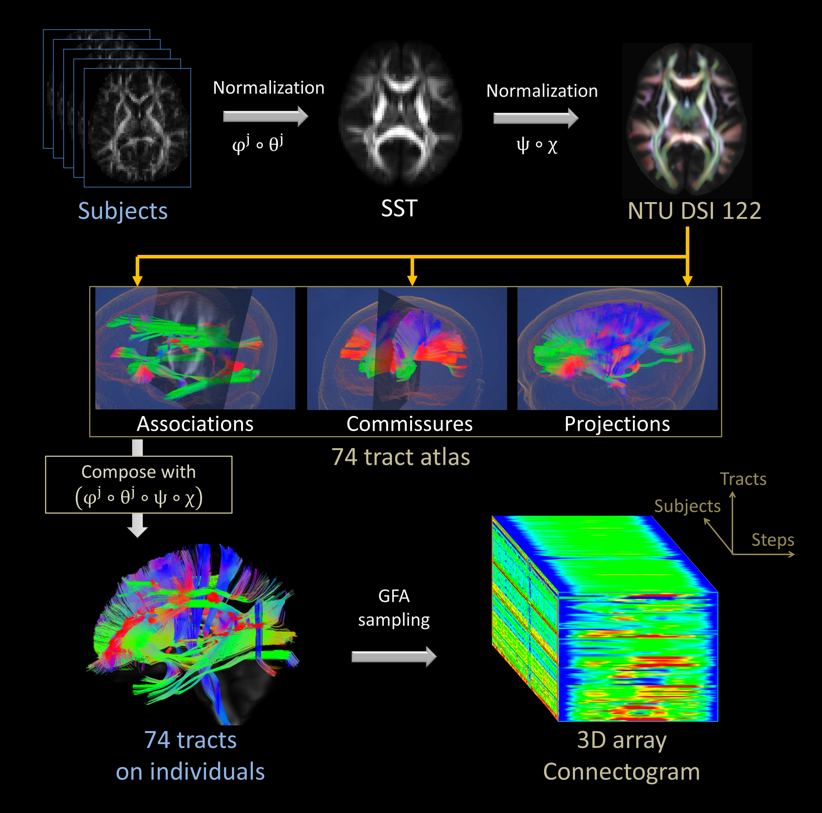
Figure 2. Tract-based automatic analysis allows automatic registration of brain images and measuring connection integrity along each tract

2016-12-21 03:44:50 / 核心實驗室

PI

Chi Kuang Sun

Chi-Kuang Sun, Distinguished Professor, Electrical Engineering, Photonics and Optoelectronics, Biomedical Electronics and Bioinformatics, National Taiwan University

Fellow IEEE, OSA, SPIE

Personal Homepage: http://ufo.ee.ntu.edu.tw/

Co-PI

Din-Ping Tsai, Distinguished Professor; Chen-Yuan Dong, Distinguished Professor; Hsuan-Shu Lee, M.D., Professor; Chen-Tung Yen, Professor; Shi-Wei Chu, Professor; Kung-Bin Sung, Associate Professor; Tzu-Ming Liu, Associate Professor; Yi-Hua Liao, M.D., Ph.D; Wei-Hsuan Yu, Assistant Professor; Wen-Jeng Lee, M.D.; Yuan Luo, Assistant Professor; Hwan-Ching Tai, Assistant Professor; Tzung-Dau Wang, Attending Physician (National Taiwan University Hospital)


THE GOALS of the optical imaging core are to improve early diagnosis and risk assessment of diseases, as well as to aid research in preventive and regenerative medicine. We aim to develop innovative optical molecular imaging technologies and establish preclinical and clinical study platforms based on these technologies by combining expertise in fundamental science, engineering and clinical research, with emphases on important health issues including cancer as well as neural, metabolic, and cardiovascular diseases.

In vivo Harmonic Generation Microscope

Optical Imaging Core In vivo Harmonic Generation Microscope Photo 01 Optical Imaging Core In vivo Harmonic Generation Microscope Photo 02
Optical Imaging Core In vivo Harmonic Generation Microscope Photo 03 Optical Imaging Core In vivo Harmonic Generation Microscope Photo 04 Optical Imaging Core In vivo Harmonic Generation Microscope Photo 05

Movable Hyperspectral Microscope System

We constructed a movable hyperspectral microscope system for acquiring spatially-resolved reflectance and fluorescence spectra. This system is used to measure oral and esophageal mucosa in vivo to quantify the scattering, absorption and fluorescence properties of the epithelium and underlying lamina propria. We evaluate the performances of the proposed method to distinguish precancerous tissue from normal tissue based on the parameters extracted from in vivo spectra.

LEICA TCS SP5 STED

2016-12-21 03:47:59 / 核心實驗室

PI

hsychen

Hsien-Yeh Chen, Professor, Department of Chemical Engineering, National Taiwan University

Co-PI

Wei-Fang Su, Distinguished Professor; Chung-May Yang M.D.and Professor; Ta-Ching Chen M.D.; Min-Huey Chen, Professor; King-Fu Lin, Professor; Wen-Bin Liau, Professor; Feng-Yu Tsai, Associate Professor; Chi-Yang Chao, Associate Professor; Jiaching Yu, Assistant Professor


The objective of molecular engineering core lab is to develop novel scaffold materials for tissue engineering through molecular design. The focus areas are in preventive and regenerative medicine in visual neural pathway and central nervous system. Standing on the basis of improving optical imaging techniques in ophthalmology, further explorations in molecular imaging are anticipated for monitoring potential therapies. We aim to achieve the goal of neural protection and regeneration in these fields by combining expertise in fundamental science, engineering and clinical research through tissue engineering technology.

Molecular Engineering Core Photo 01

Figure 1. Graging Incidence Small-Angle X-ray Scattering, GISAXS(NANOSTAR/BRUKER AXS GmbH) for morphology study of soft matter

Molecular Engineering Core Photo 02

Figure 2. Scanning near field optical microscope/confocal microscope/scanning Raman microscope(Witec alpha300, Germany) for structure and morphology study of soft matter

Molecular Engineering Core Photo 03

Figure 3. Surgical microscope and real-time optical/fluorescence imaging system for animal study(Zeiss, OPMI series, Germany)

2016-12-21 03:47:38 / 核心實驗室

PI

Ling Wei Xin

Ling-Wei Hsin, Associate Professor, School of Pharmacy, National Taiwan University

CO-PI

Ming-Fu Chang, Professor; Fu-Hsiung Chang, Professor; Min-Huei Chen, Professor; Wei-Fang Su, Distinguished Professor; Jiashing Yu, Assistant Professor


The mission of Molecular Probes Development Core is to discover and develop novel molecular probes for various molecular imaging technologies, especially positron emission tomography(PET), as tools for basic research, clinical diagnosis, and drug discovery. Our goals include: 1) discovery and development of molecular probes for novel biological targets as potential PET imaging agents; and 2) design and synthesis of appropriate precursors for radiosynthesis of PET raiopharmaceuticals in Positron Emission Tomography Core.

The research facility of Molecular Probes Development Core is located on the 6th floor of Center of Genomic Medicine Building(CZ18) in college of medicine and equipped with the equipment for organic synthesis(e.g., microwave synthesizer, Figure 1.), pharmaceutical analysis(e.g., high-resolution NMR and HPLC, Figure 2. and Figure 3.), and drug discovery and development(e.g., a fully automated instrument for measurement of various physicochemical properties of candidate compounds, Figure 4.).

Molecular Probes Development Core Photo 01

Figure 1. CEM Discovery microwave synthesizer

Molecular Probes Development Core Photo 02

Figure 2. Analytical and preparative HPLCs

Current research projects in Molecular Probes Development Core include: 1) design and synthesis of 11C- and 18F-labeled molecular imaging agents for P-glycoprotein(P-gp, ABCB1) expression and activity; 2) development of tau protein molecular probes for early diagnosis of Alzheimer's disease; 3) evaluation of 18F-labeled radiopharmaceuticals for diagnosis of metastasis of pancreatic tumor; 4) design and synthesis of 11C- and 18F-labeled molecular imaging agents for serotonin type 7(5-HT7) receptor; and 5) development of novel 18F-labeled molecular probes for dopamine transporter(DAT) and serotonin transporter(SERT).

Molecular Probes Development Core Photo 03

Figure 3. BRUKER AVANCE 600 AV nuclear magnetic resonance(NMR) spectrometer

Molecular Probes Development Core Photo 04

Figure 4. Sirius T3. A fully automated instrument for "gold standard" measurement of pKa, log P/D, and solubility using milligram sample quantities

2016-12-21 03:46:55 / 核心實驗室

PI

Ruo Fang Yan

Ruo-Fang Yan, Director, Department of Nuclear Medicine, National Taiwan University Hospital

Co-PI

Ling-Wei Hsin, Associate Professor; Chun-Jung Lin, Associate Professor; Lih-Chu Chiou, Professor; Wei-Chung Hsu, Assistant Professor; Tong-Rong Jan, Professor; Chien-Hung Chen, Associate Professor; Wen-Ming Hsu, Associate Professor; Chih-Hung Hsu, Assistant Professor; Ming-Jang Chiu, Associate Professor; Wuh-Liang Hwu, Professor; Chyng-Yann Shiue, Professor


The goal of the PET core is to use various PET probes in the study of molecular characteristics of biology, physiology, pathology and disease status of the biological systems from small animals to human beings by PET imaging. This non-invasive molecular imaging technique is useful in diagnosis, staging, monitoring and follow up of diseases related to malignancies, degenerative neuronal diseases and ischemic cardiovascular disorders.

Our Core is equipped with two PET/CT scanners for human studies, one PET/CT scanner for small animal research and one cyclotron with full capability of radiopharmaceutical synthesis. Since 2005, the production of PET probes in our lab beyond 18FDG outnumbered almost all other PET Centers in Taiwan.

Hepatocellular carcinoma(HCC) and Alzheimer's Dementia(AD) are now the two main diseases of interest in our lab. We are now performing a human clinical trial in applying 11C-PIB in AD and a multicenter clinical trial in applying 18F-choline in HCC. Furthermore, we started year 2013 using multiple PET probes, 18FDGal and 18F-choline, for the diagnosis of patients with HCC.

Positron Emission Tomography Core Photo 01

Figure 1. PET/CT scanner

Positron Emission Tomography Core Photo 02

Figure 2. Small animal PET/CT scanner

Positron Emission Tomography Core Photo 03

Figure 3. 3D 11C-PiB distribution in healthy control(above) and AD(below)

We use two PET probes(18FDOPA AND 18F-fallypride) and one SPECT probe(99mTc-TRODAT-1) to characterize children with congenital AADC deficiency preparing for gene therapy. The images showed low AADC enzyme content, intact dopamine transporter and intact dopamine D2 receptor. The post therapy image demonstrated increased AADC enzyme effect.

Positron Emission Tomography Core Photo 04

Figure 4. 18F-DOPA before Tx 99mTc-TRODAT 18F-Fallypride 18F-DOPA post Tx

In the era of molecular imaging, we can use several nuclear, both SPECT and PET, probes to target different molecules to define the biochemical phenotypes of diseases. This is true in diseases, such as neuroblastoma, hepatocellular carcinoma, Parkinson's disease, and Alzheimer's dementia. The multiple faces of a disease can be clearly defined using this imaging technique.

2016-12-21 03:45:20 / 核心實驗室

PI

Zhi Hong Chen

Jyh-Horng Chen, Professor, Department of Electrical Engineering, National Taiwan University

Co-PI

Hong-Chang Yang, Professor; Yeun-Chung Chang, Professor; Ang Yun, Clinical Associate Professor; Chia-Hsien Cheng, Associate Professor


In these five years, the aims of MRI core lab are

Develop Wideband sequence with fast, or high spatial resolution MR imaging for early detection of diseases
Design on quantitative susceptibility MR molecular imaging mapping for longitudinal studies of animals/patients
Develop DCE MRI for assessment of lung cancer mice model in variable of drugs and treatment protocol
Work on Translational Medicine on newly developed novel molecular imaging protocols for early detection of cancers

Current Studies

1. Wideband(WB) MRI

Wideband MRI is a technique that utilized wide bandwidth in order to increase the information obtained per unit time. It has the ability to speedup various applications especially those that require large coverage. In other cases, it could be also used for higher spatial resolution or temporal resolution for possible detection of diseases. The versatility of Wideband MRI allows its acceleration ability to be applied to several MRI sequences by means of echo planar imaging, perfusion, image flow, angiography, temperature,T1 imaging, T2 imaging, diffusion and the like.

Magnetic Resonance Imaging Core Photo 01

Figure 1. Conceptual illustration of a Wideband MRI human whole body scan with Wideband factor W=5

Magnetic Resonance Imaging Core Photo 02

Figure 2. The upper raw: the rat brain diffusion image in resolution of 180 x 180 um, and the bottom raw in high resolution 120 x 120 um using Wideband diffusion technique in the same scanning time

2. Quantitative susceptibility mapping (QSM)

QSM aims to quantify the susceptibility within tissues. Our study proposed a novel methodology for noninvasively detecting cerebral small vessels including veins and venules, called QSM-mMRA. The QSM-mMRA can simultaneously provide the information on cerebral anatomy, micro-vascular architecture, and SvO2, which can be used to evaluate the physiological and functional characteristics of microvascular changes for longitudinal monitoring and therapeutic evaluating with disease model.

Magnetic Resonance Imaging Core Photo 03

Figure 3. Quantitative visualisation of QSM of a normal rat brain in three orthogonal views. Veins in cortical and internal brain are indicated

Magnetic Resonance Imaging Core Photo 04

Figure 4. Detection of the rehabilitation of a cortical venule in the rat brain after stroke

Facility

Magnetic Resonance Imaging Core Photo 05

A. Siemens Prisma 3T Human MRI

B. Bruker 3T Human MRI

C. Bruker Biospec 7T Animal MRI

2016-12-21 03:46:13 / 核心實驗室

PI

Zhong Ming Chen

Chung-Ming Chen, Professor, Institute of Biomedical Engineering, National Taiwan University

Co-PI

Chiun-Sheng Huang, Professor; Tung-Wu Lu, Professor


The uniqueness of the IR imaging lies in its capability in revealing such metabolic information as heat, oxygenation, and so on, which may not be easily provided by other medical imaging modalities. With the unique strength of the IR imaging, the IR imaging core aims to develop novel low-cost, non-invasive and hazardless multi-spectrum IR imaging technologies to offer effective alternatives to characterize and detect the anatomic and functional disorders of various diseases and changes in tumor growth and treatment response.

A Quantitative Dual-Spectrum IR(QDS-IR) System for Early Detection and Monitoring Chemotherapy Response of Breast Cancer

The QDS-IR provides new tissue parameters characterizing the functional changes in tumor growth and chemotherapy response. The unique idea is to decompose the heat energy on the body surface into those from the high- and normal-temperature tissues with the environmental and physiological influences minimized. With 60 subjects, the [test] of QDS-IR is shown to have the similar variation tendency as the SUVmax of FDG-PET in more than 90% of subjects.

QDS-IR Spectrogram Imaging System

Infrared Imaging Core Photo 01

Figure 1. The system architecture of QDS-IR

Infrared Imaging Core Photo 02

Figure 2. The dual-spectrum IR camera

A Touch-free Infrared thermographic system for Real-time Remote-Monitoring of Free Flap Pedicle Thrombosis

To detect the free flap pedicle thrombosis, a new technique based on the Structural Equation Modeling(SEM) has been developed to reveal the temperature change by eliminating the effect of the latent factors. For the clinical study, the SEM-retrieved specific temperature of the flap demonstrates an evident temperature drop 80 minutes earlier than the clinical finding.

An IR Descriptor for Quantifying The Treatment Response of Complicated Skin And Soft Tissue Infection(cSSTI)

An effective descriptor, denoted by [test], has been proposed for treatment response assessment of cSSTI using IR Images. The uniqueness of the [test] lies in its capability to reveal the relative responses along the course of the treatment. Furthermore, the variation of the [test] was found to be closely related to the phenotype of the MR imaging in the course of cSSTI treatment.

Infrared Imaging Core Photo 03

Figure 3. The algorithmic flowchart of the QDS-IR system

Infrared Imaging Core Photo 04

Figure 4. Automatic identification of those regions with different qH tendency along breast chemotherapy

Vessel Thrombosis

Infrared Imaging Core Photo 05

Figure 5. The photos and IR images of the free flap regions of an oral cavity cancer patient and a swine

2016-12-21 03:45:49 / 核心實驗室

PI

Bai Qi Li

Pai-Chi Li, Distinguished Professor, Institute of Biomedical Electronics and Bioinformatics, National Taiwan University

Co-PI

Chi-Kuang Sun, Distinguished Professor


This ultrasound core lab aims at providing ultrasound related molecular imaging services. The specific items include:

Ultrasound and photoacoustic molecular imaging: imaging services, molecular probe designs, in vitro and in vivo experiments. Using our high frequency ultrasound system with a 20MHz push probe and a 40MHz imaging probe, the liver stiffness was measured serially in C57/BL6 male mice for 4 weeks by shear wave elasticity.

HFU System

 Ultrasound Imaging Core Photo 01  Ultrasound Imaging Core Photo 02  Ultrasound Imaging Core Photo 03

Custom design ultrasound sensors: leveraging existing equipments, providing custom design services of ultrasound sensors, including ceramic sensors, MEMS sensors and theragnostic sensors.

 Ultrasound Imaging Core Photo 04  Ultrasound Imaging Core Photo 05

 Ultrasound Imaging Core Photo 06

Figure 1. Serial changes of tissues elastography

For optimizing the ultrasound image-guided injection performance, a unique needle injector has been created to integrate with the probe of the high frequency ultrasound imaging system. The image plane is always coplanar with the needle. The needle image can be seen during the whole injection process.